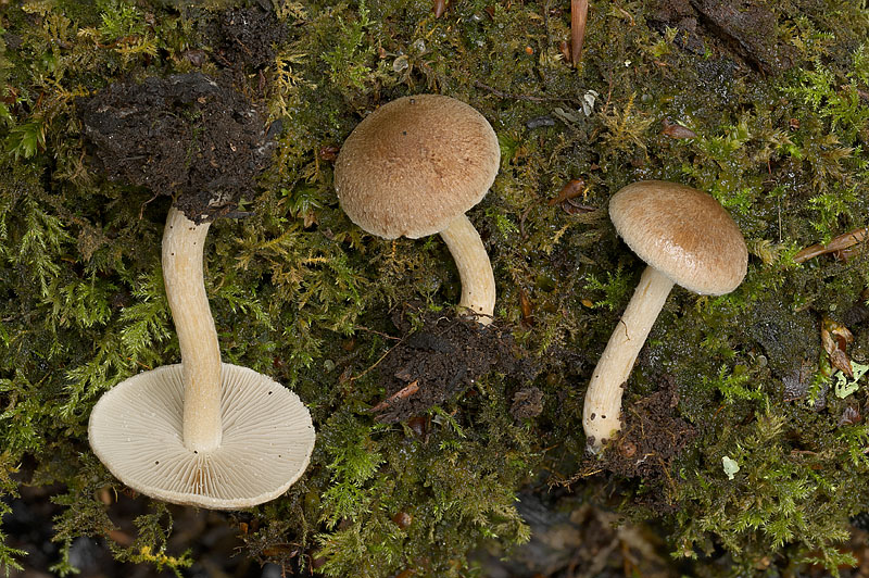
20 September 2025 Near Lyndhurst, New Forest, Hampshire. Photograph copyright Leif Goodwin. Cap Initially convex and connected at the margin to the stem by a fine veil, then expanding with a central bump, surface fibrous, brown, breaking up into scales to reveal the lighter flesh below, veil remnants visible at the margin, to about 5 cm across Gills Adnexed, crowded, pale brown then cinnamon, with a white edge Stem Cylindrical, white then brown, bruising brown, finely hairy at the apex Flesh Whitish Smell Faint, bleach Taste Do not taste Season Late summer to autumn Distribution Infrequent Habitat Usually in deciduous woods, rarely with conifers Spore Print Snuff brown Microscopic Features Spores almond shaped, smooth (8-11) x (4.5-6) µm2. Basidia club shaped, four spored. Gill edge cystidia flask shaped and spindle shaped with apical crystals. Cystida at the stem apex finger shaped. Edibility Poisonous 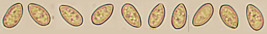
Spores in Congo Red solution viewed with a 100X immersion objective. 20 September 2025 Near Lyndhurst, New Forest, Hampshire. Photograph copyright Leif Goodwin. 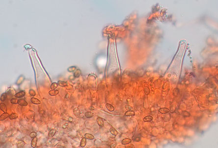
Gill edge cystidia in Congo Red solution viewed with a 40X objective. 20 September 2025 Near Lyndhurst, New Forest, Hampshire. Photograph copyright Leif Goodwin. 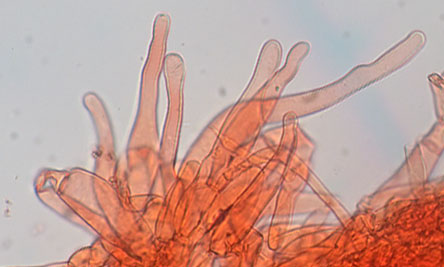
Stem cystidia in Congo Red solution viewed with a 40X objective. 20 September 2025 Near Lyndhurst, New Forest, Hampshire. Photograph copyright Leif Goodwin. 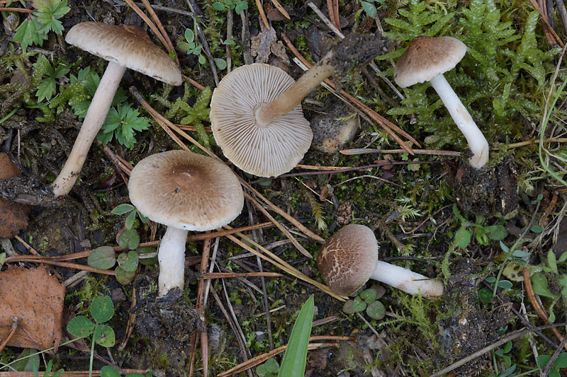
1 October 2020 Hampshire. Photograph copyright Leif Goodwin. 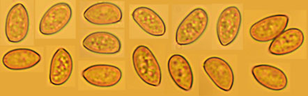
Spores in Melzer's solution viewed with a 100X immersion objective. 1 October 2020 Hampshire. Photograph copyright Leif Goodwin. 
A cystidium from the gill edge in Congo Red solution viewed with a 40X objective. 1 October 2020 Hampshire. Photograph copyright Leif Goodwin. 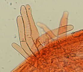
Cystidia from the stem apex in Congo Red solution viewed with a 40X objective. 1 October 2020 Hampshire. Photograph copyright Leif Goodwin. |